- Intuitive user interface
-
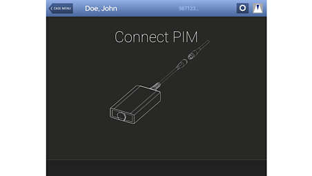
Intuitive user interface
The Core M2 system features an intuitive interface for optimal ease of use as well as guided workflows and uniform controls to simplify staff training. - Sterile field control
-
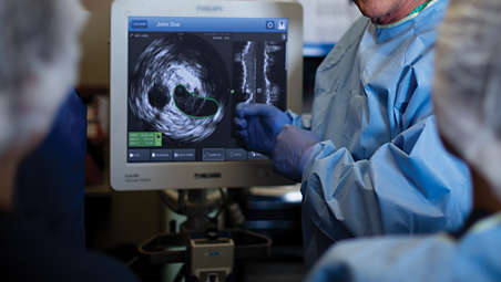
Sterile field control
The Core M2 system features a large touch screen for sterile field control, with the ability to drive from the sterile field. Only Philips offers the plug-and-play simplicity of digital IVUS and touchscreen control from the sterile field to get to your information faster. - IVUS helps with disease assessment
-
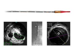
IVUS helps with disease assessment
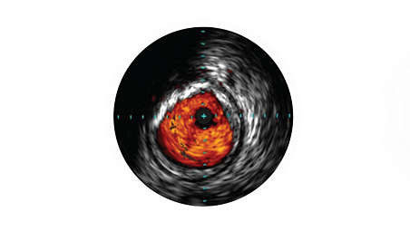
IVUS imaging helps physicians assess disease markers including plaque burden percentage, lesion location and morphology, calcium volume, and the presence of thrombus. It also enables analysis of crucial parameters – like luminal cross-sectional measurements – and helps aid in disease diagnosis. - Grayscale enhances procedures
-
Grayscale enhances procedures
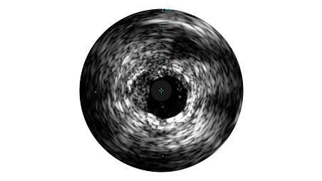
Grayscale enhances angiography procedures by enabling detailed views. Angiography produces a shadowgram of contrast, while IVUS visualizes extent and location of plaque, enabling precise disease assessment, vessel and optimal stent placement. IVUS guidance has been associated with a 74% change in PCI strategy and reduced MACE, MI, TLR, and death in large studies.¹, ² - ChromaFlo stent apposition assessment
-
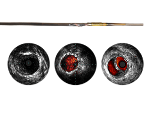
ChromaFlo stent apposition assessment
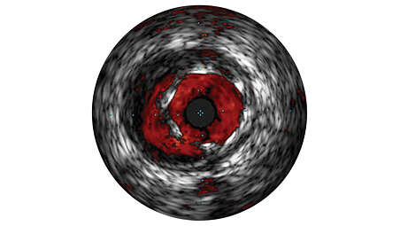
ChromaFlo highlights blood flow red for easy assessment of stent apposition, lumen size, and more. Appropriate for peripheral vessels, including, superficial femoral artery and iliac artery. It is designed to make lumen size and stent apposition instantly recognizable and helps identify branches, dissections, and plaque.
Intuitive user interface
Intuitive user interface

Intuitive user interface
Sterile field control
Sterile field control

Sterile field control
IVUS helps with disease assessment
IVUS helps with disease assessment

IVUS helps with disease assessment
Grayscale enhances procedures
Grayscale enhances procedures

Grayscale enhances procedures
ChromaFlo stent apposition assessment
ChromaFlo stent apposition assessment

ChromaFlo stent apposition assessment
- Intuitive user interface
- Sterile field control
- IVUS helps with disease assessment
- Grayscale enhances procedures
- Intuitive user interface
-
Intuitive user interface
The Core M2 system features an intuitive interface for optimal ease of use as well as guided workflows and uniform controls to simplify staff training. - Sterile field control
-
Sterile field control
The Core M2 system features a large touch screen for sterile field control, with the ability to drive from the sterile field. Only Philips offers the plug-and-play simplicity of digital IVUS and touchscreen control from the sterile field to get to your information faster. - IVUS helps with disease assessment
-
IVUS helps with disease assessment
IVUS imaging helps physicians assess disease markers including plaque burden percentage, lesion location and morphology, calcium volume, and the presence of thrombus. It also enables analysis of crucial parameters – like luminal cross-sectional measurements – and helps aid in disease diagnosis. - Grayscale enhances procedures
-
Grayscale enhances procedures
Grayscale enhances angiography procedures by enabling detailed views. Angiography produces a shadowgram of contrast, while IVUS visualizes extent and location of plaque, enabling precise disease assessment, vessel and optimal stent placement. IVUS guidance has been associated with a 74% change in PCI strategy and reduced MACE, MI, TLR, and death in large studies.¹, ² - ChromaFlo stent apposition assessment
-
ChromaFlo stent apposition assessment
ChromaFlo highlights blood flow red for easy assessment of stent apposition, lumen size, and more. Appropriate for peripheral vessels, including, superficial femoral artery and iliac artery. It is designed to make lumen size and stent apposition instantly recognizable and helps identify branches, dissections, and plaque.
Intuitive user interface

Intuitive user interface

Intuitive user interface
Sterile field control

Sterile field control

Sterile field control
IVUS helps with disease assessment

IVUS helps with disease assessment

IVUS helps with disease assessment
Grayscale enhances procedures

Grayscale enhances procedures

Grayscale enhances procedures
ChromaFlo stent apposition assessment

ChromaFlo stent apposition assessment

ChromaFlo stent apposition assessment
Documentation
-
Brochure (1)
-
Brochure
- Core M2 IVUS workflow postcard (782.9 kB)
-
Technical data sheet (1)
-
Technical data sheet
- Core M2 data sheet (732.0 kB)
-
Brochure (1)
-
Brochure
- Core M2 IVUS workflow postcard (782.9 kB)
-
Technical data sheet (1)
-
Technical data sheet
- Core M2 data sheet (732.0 kB)
-
Brochure (1)
-
Brochure
- Core M2 IVUS workflow postcard (782.9 kB)
-
Technical data sheet (1)
-
Technical data sheet
- Core M2 data sheet (732.0 kB)
Teknik özellikler
- Power requirements
-
Power requirements System input - 100-240VAC +50/50Hz, 200W
Workstation - 100-240VAC +50/50Hz, 200W
-
- Dimensions
-
Dimensions Panel PC - H=15.75", W=18", D=3.13"
Core M2 cart - H=58.75", W=26.25", D=26"
-
- Ordering information
-
Ordering information Core M2 vascular system - 400-0100.17
SA PIM - 802143001-ROHS
Cart - 400-0100.18
Snap Kovers, 28” x 20 (optional) - 01-2820 (send orders to orders@advmeddes.com)
-
- Power requirements
-
Power requirements System input - 100-240VAC +50/50Hz, 200W
Workstation - 100-240VAC +50/50Hz, 200W
-
- Dimensions
-
Dimensions Panel PC - H=15.75", W=18", D=3.13"
Core M2 cart - H=58.75", W=26.25", D=26"
-
- Power requirements
-
Power requirements System input - 100-240VAC +50/50Hz, 200W
Workstation - 100-240VAC +50/50Hz, 200W
-
- Dimensions
-
Dimensions Panel PC - H=15.75", W=18", D=3.13"
Core M2 cart - H=58.75", W=26.25", D=26"
-
- Ordering information
-
Ordering information Core M2 vascular system - 400-0100.17
SA PIM - 802143001-ROHS
Cart - 400-0100.18
Snap Kovers, 28” x 20 (optional) - 01-2820 (send orders to orders@advmeddes.com)
-
Related products
Alternative products
-
Visions PV .035
- Digital IVUS catheter evaluates vascular morphology in blood vessels
- Provides cross-sectional imaging of these vessels
- 90 cm length and 60 mm max imaging diameter for 0.035” guide wire interventional procedures
- Grayscale IVUS capable
Ürünü görüntüle
-
Visions PV .018
- Digital IVUS catheter evaluates vascular morphology in blood vessels
- Provides cross-sectional imaging of these vessels
- 135 cm working length and 24 mm max imaging diameter for 0.018” guide wire interventional procedures
- Grayscale IVUS and ChromaFlo capable
Ürünü görüntüle
-
Visions PV .035
As an adjunct to conventional angiographic interventions, the Visions PV .035 digital IVUS catheter evaluates vascular morphology in blood vessels and provides cross-sectional imaging of these vessels. With a 90 cm length and 60 mm max imaging diameter for 0.035” guide wire interventional procedures, the device aids in peripheral artery disease diagnosis and venous disease and guides clinicians toward the correct therapy for the patient’s unique needs.
Ürünü görüntüle
-
Visions PV .018
As an adjunct to conventional angiographic interventions, the Visions PV .018 digital IVUS catheter evaluates vascular morphology in blood vessels and provides cross-sectional imaging of these vessels. With a 135 cm working length and 24 mm max imaging diameter for 0.018” guide wire interventional procedures, the device aids in peripheral artery disease diagnosis and guides clinicians toward the correct therapy for the patient’s unique needs.
Ürünü görüntüle
- 1. Witzenbichler B et al. Relationship Between Intravascular Ultrasound Guidance and Clinical Outcomes After Drug-Eluting Stents: The ADAPT-DES Study. Circulation 2014 Jan: 129,4;463-470
- 2. Ahn et al. Meta-Analysis of Outcomes After Intravascular Ultrasound Guided Versus Angiography-Guided Drug-Eluting Stent Implantation in 26,503 Patients Enrolled in Three Randomized Trials and 14 Observational Studies. Am J Cardiol 2014; 113:1338-1347
- * Product availability is subject to country regulatory clearance. Please contact your local sales representative to check the availability in your country.
- Always read the label and follow the directions for use.
- Philips medical devices should only be used by physicians and teams trained in interventional techniques, including training in the use of this device.
- Philips reserves the right to change product specifications without prior notification.
- ©2025 Koniklijke Philips N.V. All rights reserved. Trademarks are the property of Koninklijke Philips N.V. or their respective owners.
