Magnetic Resonance
MR-only simulation  Unleash the real power of MR simulation
Bu ürün artık mevcut değil

MR-only simulation

Unleash the real power of MR simulation

Bu ürün artık mevcut değil

Benzer ürünler bul

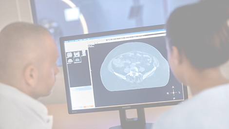
Innovative MR-only simulation pelvis lets you plan radiation therapy for male and female pelvic cancer patients with soft-tissue tumors using MRI as a single-modality solution. Within just one MR exam, MR-only simulation provides excellent soft-tissue contrast for target and OAR delineation, and CT-like density information for dose calculations. This not only extends the benefits of MRI’s outstanding soft-tissue contrast to radiotherapy planning, but it also eliminates arduous, error-prone CT-MRI registration from the process, reducing uncertainties and complexity.
Teknik özellikler

MR-only simulation pelvis
Anatomies
  • Male and female pelvis, which includes prostate, bladder, rectum, anus and cervix
Compatibility RTdrive
  • No
Compatibility with MR-based auto-contouring
  • No
Density maps
  • Continuous Hounsfield Units
MR-only simulation prostate
Anatomies
  • Prostate
Compatibility RTdrive
  • Yes
Compatibility with MR-based auto-contouring
  • Yes, for prostate and prostate OARs
Density maps
  • Discrete, five different classifications: water, fat, spongy bone, cortical bone, and air
MR-only simulation
MR-only simulation
Compatibility
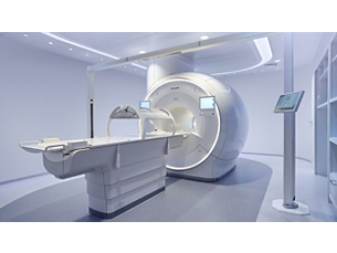
  • Ingenia MR-RT 1.5T and 3.0T
Imaging protocol
  • Dedicated ExamCard provides sequences for:
  • 3D MRCAT source information (mDIXON)
  • Anatomical information for delineation (T2W TSE)
MRCAT generation
  • Running parallel to image acquisition on the MR console, embedded post-processing generates MRCAT images using:
  • Automated segmentation and tissue classification
  • Automated assignment of CT-based density values
Export to treatment planning systems
  • MRCAT images are DICOM conform (CT) for direct export to treatment planning systems
MR-only simulation pelvis
Anatomies
  • Male and female pelvis, which includes prostate, bladder, rectum, anus and cervix
Compatibility RTdrive
  • No
MR-only simulation prostate
Anatomies
  • Prostate
Compatibility RTdrive
  • Yes
See all specifications
MR-only simulation pelvis
Anatomies
  • Male and female pelvis, which includes prostate, bladder, rectum, anus and cervix
Compatibility RTdrive
  • No
Compatibility with MR-based auto-contouring
  • No
Density maps
  • Continuous Hounsfield Units
MR-only simulation prostate
Anatomies
  • Prostate
Compatibility RTdrive
  • Yes
Compatibility with MR-based auto-contouring
  • Yes, for prostate and prostate OARs
Density maps
  • Discrete, five different classifications: water, fat, spongy bone, cortical bone, and air
MR-only simulation
MR-only simulation
Compatibility
  • Ingenia MR-RT 1.5T and 3.0T
Imaging protocol
  • Dedicated ExamCard provides sequences for:
  • 3D MRCAT source information (mDIXON)
  • Anatomical information for delineation (T2W TSE)
MRCAT generation
  • Running parallel to image acquisition on the MR console, embedded post-processing generates MRCAT images using:
  • Automated segmentation and tissue classification
  • Automated assignment of CT-based density values
Export to treatment planning systems
  • MRCAT images are DICOM conform (CT) for direct export to treatment planning systems
  • 1. Accuracy means: MRCAT provides around 1 mm total geometric accuracy of image data in less than 20 cm Diameter Spherical Volume (DSV). MRCAT provides below 2 mm total geometric accuracy of image data in more than 40 cm Diameter Spherical Volume (DSV)*. * Limited to 32 cm in z-direction in more than 95% of the points within the volume
  • 2. Equivalent means: the simulated dose based on MRCAT images does not differ in 95% of the pelvic cancer patients (Gamma analysis criterion 3%/3mm realized in 99% of voxels within the PTV or exceeding 75% of the maximum dose) when compared with CT-based plan for EBRT.
  • 3. Tested in a non-clinical environment with single Pinnacle user and a 5-beam IMRT plan. Excluding time for optional manual adjustments.

You are about to visit a Philips global content page

Continue

Sitemiz Microsoft Edge, Google Chrome veya Firefox'un en son sürümüyle en iyi şekilde görüntülenebilir.