FieldStrength MRI magazine
User experiences - February 2019
Share this article:
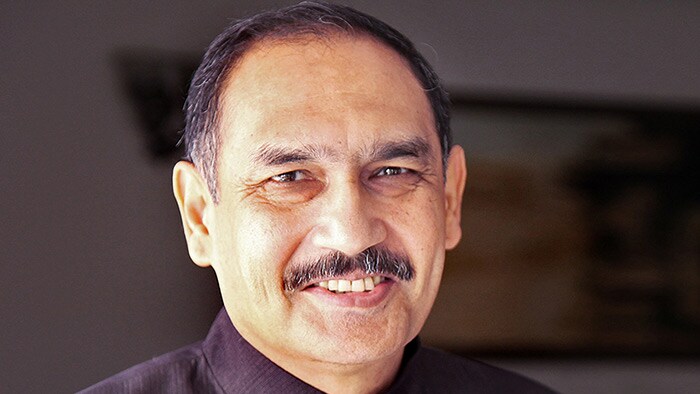
Harsh Mahajan, MD, Founder of Mahajan Imaging, a chain of leading imaging centers in New Delhi, India. He is Chief Radiologist and has over 30 years of experience in MRI.
Combining economic soundness and clinical performance in MRI with Prodiva 1.5T
When choosing the Ingenia Prodiva 1.5T scanner for two of his practices in relatively smaller hospitals (<200 beds), Dr. Harsh Mahajan had to find a balance between having state-of-the-art, future-proof imaging capabilities, and spending only a reasonable amount of money. After about a year of operation, Dr. Mahajan is highly satisfied with the Prodiva system, which not only brought savings in installation and running cost, but also excellent clinical capabilities.
“With Prodiva we were getting a high-end system at a reasonable price, and because it includes latest technology I expect a long lifetime of the system”
Versatility coupled with cost savings
Mahajan Imaging, based in New Delhi, India, acquired two Ingenia Prodiva 1.5T MRI machines in 2017. By choosing the Prodiva, Mahajan Imaging realized several cost savings associated with installation, scanning procedures, and power usage. Prodiva’s capabilities include both basic and advanced applications for neurology, body, musculoskeletal (MSK), oncology, and cardiovascular imaging. The design of the machine is lightweight and compact, allowing for easy siting and less need for renovation. The staff was pleasantly surprised by the versatility of the machines and the quality of the images.
In 2017, Mahajan Imaging acquired two Prodiva 1.5T scanners at two medium-sized private hospitals located in Delhi: Fortis Hospital and PSRI Hospital. Dr. Mahajan says “Our preference was for a scanner with the capability to perform high-end applications and originally we thought we needed a 3 Tesla machine for that. However, we knew that a 3 Tesla system would be very expensive and we would never be able to break even, considering that in India, we only charge about the equivalent of about 100 USD for an MRI scan.”
Proven digital technology and high-end applications
Mahajan Imaging, founded by Dr. Harsh Mahajan, is a leading chain of imaging centers in India. The company currently includes 50 radiologists and other physicians and 350 support
So when Dr. Mahajan started looking for novel technology in the 1.5T range that was able to produce
“We were able to negotiate the Prodiva through the narrow corridors of the hospital without having to break down any walls or remove any ceiling”

Versatility coupled with cost savings
Mahajan Imaging, based in New Delhi, India, acquired two Ingenia Prodiva 1.5T MRI machines in 2017. By choosing the Prodiva, Mahajan Imaging realized several cost savings associated with installation, scanning procedures, and power usage. Prodiva’s capabilities include both basic and advanced applications for neurology, body, musculoskeletal (MSK), oncology, and cardiovascular imaging. The design of the machine is lightweight and compact, allowing for easy siting and less need for renovation. The staff was pleasantly surprised by the versatility of the machines and the quality of the images.
Mahajan Imaging, founded by Dr. Harsh Mahajan, is a leading chain of imaging centers in India. The company currently includes 50 radiologists and other physicians and 350 support staff, and encompasses more than 30 years of MRI experience. Mahajan Imaging runs 9 centers, including stand-alone facilities and in hospital departments. In 2017, Mahajan Imaging acquired two Prodiva 1.5T scanners at two medium-sized private hospitals located in Delhi: Fortis Hospital and PSRI Hospital. Dr. Mahajan says “Our preference was for a scanner with the capability to perform high-end applications and originally we thought we needed a 3 Tesla machine for that. However, we knew that a 3 Tesla system would be very expensive and we would never be able to break even, considering that in India, we only charge about the equivalent of about 100 USD for an MRI scan.”
Proven digital technology and high-end applications
So when Dr. Mahajan started looking for novel technology in the 1.5T range that was able to produce high quality scans, Prodiva 1.5T had just entered the market. “In essence, this was our reason: high quality imaging and the fact that this was absolutely new technology, which would not quickly become obsolete. Prodiva 1.5T is a digital system and incorporates very recent technology that would last for at least a decade, if not longer,” says Dr. Mahajan. “And when we saw the Prodiva 1.5T for the first time, we were surprised to discover that this was actually a very beautiful machine.”
“We were able to negotiate the Prodiva through the narrow corridors of the hospital without having to break down any walls or remove any ceiling”
Realized cost saving with Prodiva installation
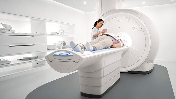
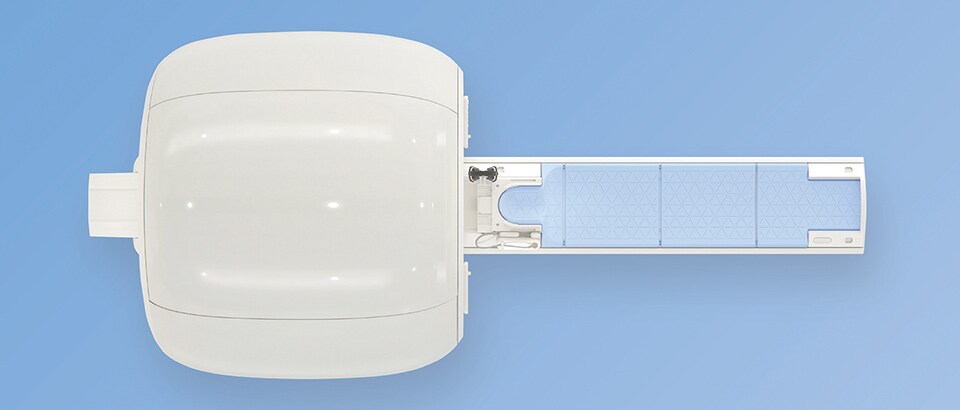
Also the compact, lightweight MRI magnet made Prodiva an attractive choice. Mahajan Imaging clearly benefitted from the compact size of the Ingenia Prodiva scanner: “Since the MRI scanners were going into already-running hospitals, originally we had foreseen having to break a lot of walls in the corridors and break the ceiling for introducing a new MRI scanner,” says Dr. Mahajan. “But fortunately, we were able to negotiate the Prodiva through the narrow corridors of the hospital into its resting place without having to break down any walls or remove any ceiling! According to our estimate, we saved approximately 20,000 USD on wall reconstruction and 30,000 USD on ceiling renovations – damage which would have been needed for a larger machine. There would have been business lost during the renovations as well, which was saved now.” Prodiva’s small footprint allows housing it where other MRI scanners may not fit. It features a small fringe field and only weighs only 2900 kg. “The machine requires much less space, so the MRI suite can be much smaller, leaving space for other equipment. Most importantly, I didn’t have to worry about reinforcing the floor on which the magnet would be housed; this would not only have been very costly, but also very cumbersome. Our siting costs were lower than originally foreseen.”

Long-term cost control with Prodiva
In addition to installation cost, also the longevity, reliability and lifetime cost of the MRI were factors influencing Dr. Mahajan’s purchase decision. “With Prodiva 1.5T we were getting a highend system at a reasonable price, and because it includes latest technology I expect a long lifetime of the system,” he says. “The low power use of Prodiva helps to keep our energy cost low. We have already experienced that the power requirement is much less than it is for other MRI scanners. We used to need up to 120 kVA, but for Prodiva this number is down to 50 kVA, so even there we find cost savings.” Dr. Mahajan notes that “some of the other major considerations are the quality of the service and the frequencies of breakdowns.” He and his staff appreciate the high amount of uptime they experience with the Prodiva system. “There have haven’t been any significant breakdowns in the past 16 months since we have had the machine, so we never had to send a patient away because the machine was down.” The flexibility gained by using the Prodiva flexible phased array MSK coils also brings down the price of imaging, according to Dr. Mahajan. Previously, each had a dedicated coil. “I have always bought dedicated coils for every joint and body part, which is no longer necessary. Prodiva’s smaller digital MSK coils can be combined to become larger coils. By fitting them together, we can image both small and large body parts using the same equipment.”
“The low power use of Prodiva helps to keep our energy cost low”
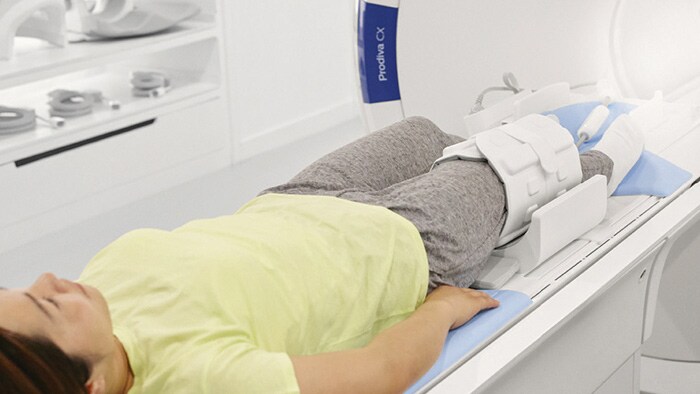
“Prodiva’s smaller digital MSK coils can be combined to become larger coils”
Flexible coils eliminate restrictions in coil orientation
“The flexible digital MSK coils felt like a revolution: we can use them in any plane we want: longitudinal, horizontally, or vertically, and they function,” says Dr. Mahajan. “These flexible coils allow for imaging a smaller FOV with one coil, and larger FOV with multiple coils in tandem. In some exams, for instance thigh and leg, techs don’t need to move the coils during scanning anymore, which offers a versatility which was never possible with any other coils. Use of these coils also allows for whole body imaging, with imaging coverage of 190 cm with 50 x 50 x 45 cm field of view (FOV).”
Dr. Mahajan mentioned that he did not work with circular flex coils before he had Prodiva. “Now, we not only we use the circular coil for small body parts, but by putting two of the circular coils into a Positioner for doing MR mammography, we actually create a 14-channel mammography coil, and the image quality is phenomenal.”
“The shorter set-up times for the patients and coils are much appreciated and scans are usually performed quickly”
Decreased patient set-up time
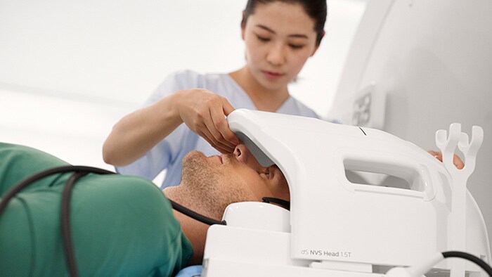
The staff at Mahajan Imaging benefits from the simplified Breeze workflow, which reduces the number of coil positioning steps and reduces patient set-up time. Although both facilities with Prodiva do not usually have large throughput requirements, the shorter set-up times for the patients and the coils are much appreciated and scans are usually performed quickly. “Another factor that helps make patient positioning very easy is the low table height of Prodiva 1.5T, that often does not need to be adjusted,” says Dr. Mahajan. “Most of our average-height patients can actually just sit on the table and lie down, and that reduces set-up time.” Working with Prodiva is easy to learn, according to Dr. Mahajan. “The system is simple to use. Our technologists were able to adapt to and use the interface very quickly. Some of our operators had never worked with Philips MRI systems previously, but adapted to it very quickly. It was easy for the operators to develop an indepth understanding of the software and the various available applications.”
Patients can appreciate the effect of Prodiva features on comfort
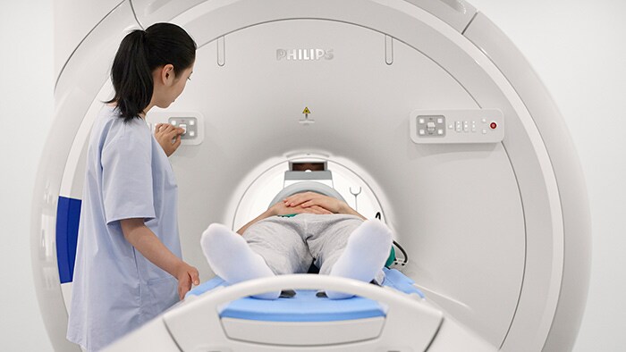
To conclude, Dr. Mahajan summarizes the factors that have contributed to his satisfaction. “Ingenia Prodiva is capable of doing very good diagnostic scanning with short set-up times. Because of that, our throughput is fluent and quicker, and our patients appreciate non-intimidating design and muted acoustic noise.” From an economic perspective, he notes “Thanks to the compact size and light weight our installation was much cheaper than anticipated and our siting costs are lower, since the area required for the machine is much smaller. The machine was reasonably priced and we did not need to purchase dedicated coils. The uptime is high and our savings on costs of electricity for example, are pretty substantial.”
Robust imaging even in presence of motion, metal or fat
Obtaining high quality imaging in a broad group of patients requires dealing with factors that may impact image quality. Prodiva offers some powerful tools to help reduce artifacts that can potentially hamper diagnostic confidence or lead to repeat scans. “Movement, however minor, creates artifacts on images. Fortunately, MultiVane XD reduces motion artifacts, which helps us very much with patients who are less cooperative or who are semi-conscious, as well as patients with stroke.” “The method that Prodiva 1.5T offers for reducing the artifacts caused by metal also works very well, both in the extremities around implants as well as in the spine. In order to obtain robust, uniform fat suppression we can use the mDIXON fat suppression, which is especially helpful in the extremities and in whole body imaging.”
“The low acoustic noise and the short bore of Prodiva can help patients feel comfortable”
“Our installation was much cheaper than anticipated and our siting costs are lower, since the area required for the machine is much smaller”
Technologists can perform advanced imaging techniques with Prodiva
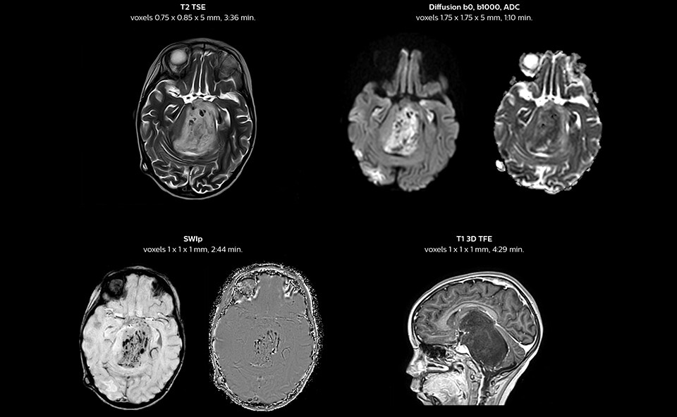
“We have been very, very pleasantly surprised by the quality of the images,” says Dr. Mahajan. “I have shown Prodiva images to people around the world during my travels.” Ingenia Prodiva 1.5T includes such features as dStream digital broadband technology to increase image quality with high SNR. “The system came with its newest sequences that allow us to perform advanced techniques like excellent diffusion in the brain and other body parts as well, spectroscopy, detailed neurography, CSF flow studies, imaging of difficult areas like skull base, tractography, DTI, and even DTI of the spinal cord – all of this is available to us.” The radiologists at Mahajan Imaging were also impressed by the quality of cardiac MRI. “We have done some MR angiography of the coronary arteries with the Prodiva and it provided excellent coronaries – and this was without using any contrast agent. We can also perform fantastic ngiography of other body parts.” “With Prodiva 1.5T, the images are very crisp and sharp, and the resolution is very good,” says Dr. Mahajan. “Imaging can be done in short scan times with excellent quality and broad range of possible applications, whether it is angiography, a scan with 3D reconstruction, any kind of more special images, what we do, the sharpness, resolution and the ease of use – everything counts in total when we are reporting.”
Enhanced clinical capabilities with a robust return on investment
To conclude, Dr. Mahajan summarizes the factors that have contributed to his satisfaction. “Ingenia Prodiva is capable of doing very good diagnostic scanning with short set-up times. Because of that, our throughput is fluent and quicker, and our patients appreciate non-intimidating design and muted acoustic noise.” From an economic perspective, he notes “Thanks to the compact size and light weight our installation was much cheaper than anticipated and our siting costs are lower, since the area required for the machine is much smaller. The machine was reasonably priced and we did not need to purchase dedicated coils. The uptime is high and our savings on costs of electricity for example, are pretty substantial.”
“In essence, we chose Prodiva 1.5T for its high quality imaging and because it was new technology, which would not quickly become obsolete”
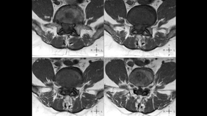
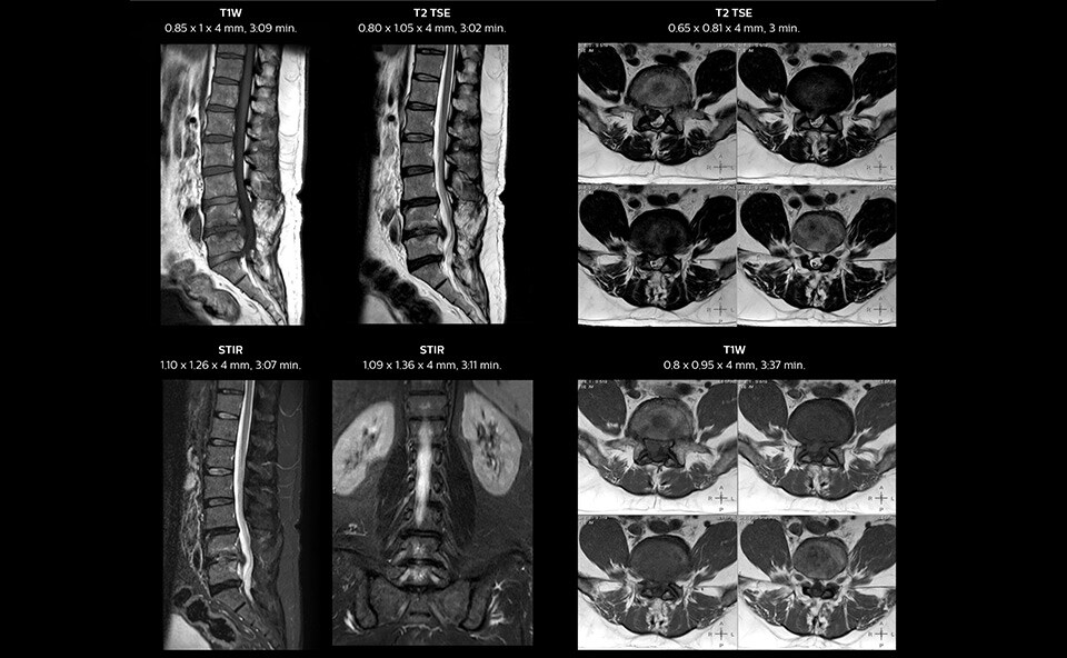
MRI of spondylosis in lumbar spine
In this patient MRI images show spondylotic degenerative changes in the lumbar spine, in view of the osteophyte, disc desiccations and Sc hmorl’s nodes with lumbarization of S1, post-laminectomy changes at L5-S1 and S1-S2 levels with disc protrusion and extrusion, and diffuse disc bulges from L3-L5 with nerve root compression.
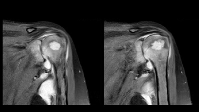
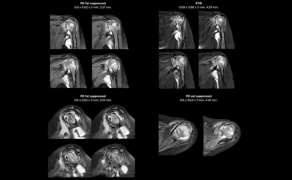
MRI of the shoulder on Ingenia Prodiva 1.5T
In these MRI images a focal lesion can be seen in the left humoral head with erosion of cortex. Multiloculated collection around the shoulder joint effusion, synovial thickening, and reduced joint space for arthritis were also suggested. The findings are compatible with infective etiology.
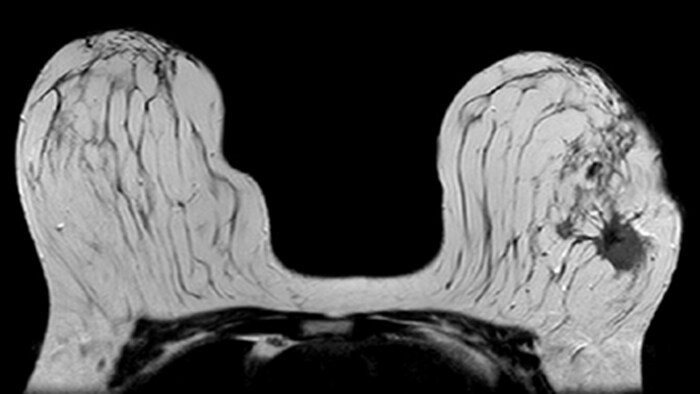
MRI of patient with invasive ductal carcinoma
In this patient, biopsy revealed invasive ductal carcinoma in the left breast (BIRADS VI). The MRI results are compatible with multifocal multicentric malignancy in the left breast, located in the upper-inner and outer quadrants, almost from the 10 to 4 o’clock position, with an extensive intraductal component, without infiltration of the underlying pectoral muscles.
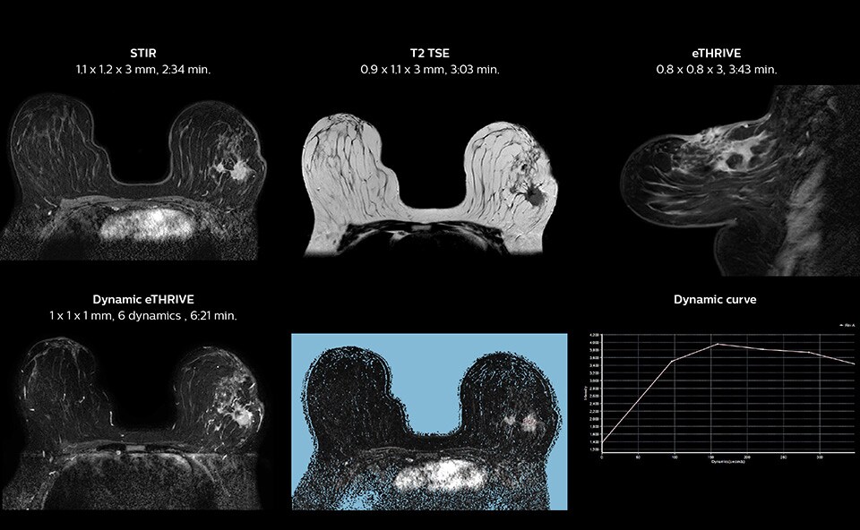
Summary of Mahajan Imaging experience with Ingenia Prodiva 1.5T:
Results from case studies are not predictive of results in other cases. Results in other cases may vary.
Share this article:
Subscribe to FieldStrength
Our periodic FieldStrength MRI newsletter provides you articles on latest trends and insights, MRI best practices, clinical cases, application tips and more. Subscribe now to receive our free FieldStrength MRI newsletter via e-mail.
Stay in touch with Philips MRI

Related information

More from FieldStrength
